
Congenital vertical talus is a condition present at birth that causes a very rigid flat foot.
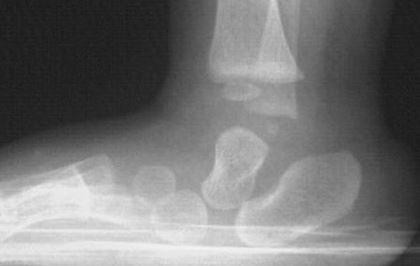
Congenital vertical talus, also known as congenital convex pes valgus or congenital rocker-bottom foot, is a rare foot deformity that is present at birth. It is characterized by a rigid and fixed dorsiflexion of the talus bone (the bone in the ankle) and a prominent convexity of the sole of the foot, resembling a rocker-bottom shape.
Symptoms: The primary symptoms of congenital vertical talus include a visible convexity of the sole of the foot, a rigid or stiff foot, and limited or absent ability to move the foot up and down. The deformity can cause pain, difficulty with walking, and may lead to associated conditions like calluses, skin irritation, or ulcers.
Causes: The exact cause of congenital vertical talus is not fully understood. However, it is believed to result from a combination of genetic and environmental factors. It can sometimes be associated with underlying conditions such as arthrogryposis, spina bifida, or genetic syndromes like trisomy 18 or trisomy 21 (Down syndrome).
Diagnosis: Congenital vertical talus is typically diagnosed shortly after birth or during infancy based on the physical appearance of the foot and limited range of motion. Imaging tests such as X-rays may be used to confirm the diagnosis and evaluate the severity of the deformity.
Treatment: The main goal of treatment for congenital vertical talus is to correct the foot deformity and improve function. Non-surgical treatment options include gentle stretching exercises, manipulation, and casting to gradually realign the foot. However, surgery is often necessary to achieve a lasting correction. Surgical procedures involve releasing tight structures, realigning the bones, and stabilizing the foot with the use of pins, screws, or plates.
Follow-up care: After treatment, regular follow-up visits with a healthcare professional, such as a pediatric orthopedic surgeon, are important to monitor the foot’s progress and address any potential issues. Physical therapy and orthotic devices may be recommended to aid in rehabilitation and support proper foot function.
The prognosis for congenital vertical talus is generally good with appropriate treatment. Early intervention is crucial to prevent complications and optimize foot function
Congenital Vertical Talus (CVT) FAQs
What is congenital vertical talus?
It is a rigid flatfoot deformity in which the talus bone points nearly straight downward (vertically) instead of its normal forward angle. The navicular bone is dorsally dislocated on the head of the talus. This produces hindfoot equinus and valgus, midfoot dorsiflexion, and forefoot abduction, creating a convex, rocker-bottom sole. One or both feet may be affected (roughly half of cases are bilateral).
How common is it?
CVT is rare, with an estimated prevalence of about 1 in 10,000 live births (some sources cite lower figures such as 1 in 150,000). It occurs in boys and girls with roughly equal frequency (or a slight male predominance in some reports).
What causes congenital vertical talus?
The exact cause is often unknown (idiopathic in more than half of cases). Roughly 50% of cases are associated with neuromuscular conditions (e.g., myelomeningocele/spina bifida, arthrogryposis), chromosomal abnormalities (e.g., trisomies 13, 15, 18), or genetic syndromes (e.g., Marfan syndrome). Rare familial cases have been linked to mutations such as in the HOXD10 gene. Other proposed factors include muscle imbalance or arrested fetal foot development.
What does it look like? What are the signs and symptoms?
- Rigid rocker-bottom sole (convex plantar surface) that cannot be manually corrected.
- Prominent talar head palpable on the medial sole.
- Hindfoot in fixed equinus and valgus; forefoot abducted and dorsiflexed.
- Deep creases on the dorsum of the foot.
- In babies it is usually painless. Untreated, older children develop pain, calluses, difficulty with shoe wear, and an abnormal gait (limited push-off, excessive heel contact).
How is it diagnosed?
Diagnosis is clinical (rigid rocker-bottom deformity noted at birth or early infancy) and confirmed with radiographs. Key views are anteroposterior and lateral foot X-rays, especially a forced plantar-flexion lateral view showing persistent dorsal dislocation of the talonavicular joint (the navicular does not reduce onto the talus). Forced dorsiflexion views help assess equinus. Doctors also evaluate for associated neuromuscular or genetic conditions, which may include additional imaging or genetic testing. It can occasionally be suspected on prenatal ultrasound.
How is congenital vertical talus different from other newborn flat feet or clubfoot?
Unlike flexible flatfoot or calcaneovalgus deformity, CVT is rigid and does not correct with gentle manipulation. Oblique talus is a milder, partially reducible variant. Clubfoot (talipes equinovarus) has the opposite deformity pattern (inverted, plantar-flexed foot). Misdiagnosis is common, so specialist evaluation is important.
How is it treated?
Treatment almost always begins with serial manipulation and casting (a “reverse Ponseti” method) to stretch soft tissues and improve alignment. Most children then need a minimally invasive procedure (typically around 6–12 months of age) involving percutaneous Achilles tenotomy and pinning of the reduced talonavicular joint. More extensive soft-tissue releases are used for stiffer or residual cases. Postoperative bracing is essential to maintain correction and reduce relapse risk. Early treatment is preferred; neglected cases may later require more complex surgery (e.g., arthrodesis or talectomy as salvage).
Can it be corrected without surgery?
Serial casting alone is rarely sufficient for true CVT because the talonavicular dislocation is rigid. Casting prepares the foot and often allows a less invasive surgical correction, but surgery is usually required. Milder oblique talus may sometimes respond to non-operative management.
What is the outlook (prognosis)?
Untreated CVT leads to progressive pain, callosities, shoe problems, and disability. With timely casting plus minimally invasive surgery and consistent bracing, most children achieve a plantigrade, functional, and relatively pain-free foot. Outcomes are generally better in isolated (idiopathic) cases than in those with underlying neuromuscular or syndromic conditions. Mild residual differences (e.g., slightly smaller foot, reduced ankle motion, or calf atrophy) can occur. Relapse is possible and requires monitoring.
Will my child need lifelong treatment or special shoes?
After initial correction and a period of bracing (often full-time then nighttime for 1–2+ years), many children walk and function well in regular shoes. Ongoing orthopedic follow-up is recommended, especially during growth. Some may benefit from orthotics later if residual issues arise.
Is there a risk of recurrence or complications?
Yes—relapse can occur, particularly if bracing is not followed diligently or if an associated condition is present. Older surgical techniques carried higher risks of stiffness, overcorrection, or avascular necrosis; modern minimally invasive approaches have improved results. Neglected deformity after age 3–4 becomes harder to correct fully.